
Issue, August 2026
Newsletter
5 High-Yield Radiology Sessions You Shouldn’t Miss
A quick look at what’s new on Conceptual Radiology
Radiology preparation becomes easier when you learn concepts through images, clinical correlation and exam-focused discussions. This month, Conceptual Radiology brings you sessions covering Peripheral Artery Disease and Aortoiliac Lesions, High-Yield Radiology Spotters, FMF Preeclampsia Screening, Doppler Assessment in Fetal Hydrops, and Meniscus Tears on MRI.
These are just a few glimpses from the learning happening inside Conceptual Radiology. Here’s what you can explore — and why these sessions are worth adding to your watchlist.
In-App Learning Highlights
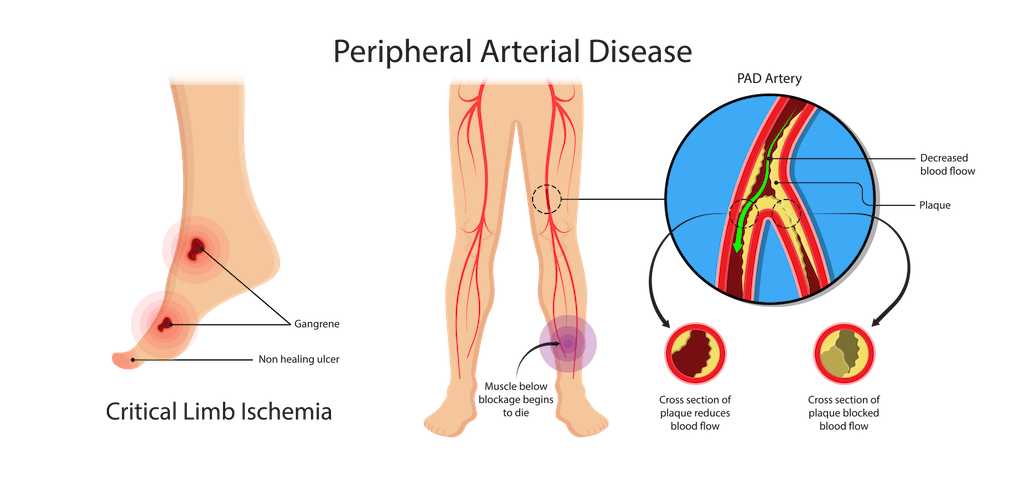
01. Peripheral Artery Disease: Diagnosis, Imaging & Aortoiliac Lesions With Dr. Akhil Monga
Peripheral Artery Disease becomes much easier to approach when you connect its clinical presentation, disease progression, imaging findings and treatment principles.
This high-yield Interventional Radiology session takes you from the basics of PAD to Aortoiliac Occlusive Disease and limb ischemia.
Key Takeaways
- Understanding Peripheral Artery Disease (PAD) & LEAD
- Atherosclerosis and disease progression
- Important risk factors and clinical presentation
- Imaging evaluation of PAD
- Aortoiliac Occlusive Disease
- Acute vs chronic limb ischemia
- Treatment principles in Interventional Radiology
- Understanding polyvascular disease
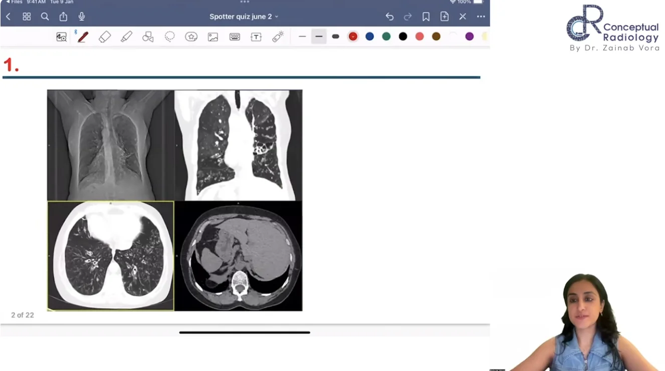
02. High-Yield Radiology Spotters for MD/DNB Residents Explained By Dr. Zainab Vora
Cartagena Syndrome, GI Lymphoma, AVN & More
Spotters are all about recognising the image, identifying the key finding and reaching the diagnosis quickly.
This session brings together several important image-based cases that can help you sharpen your practical examination preparation.
🔑 Key Takeaways
- Cartagena Syndrome / Primary Ciliary Dyskinesia
- Testicular Microlithiasis
- Striated Nephrogram in Acute Pyelonephritis
- Renal Lymphangiectasia
- Differential diagnosis of perirenal masses
- GI Lymphoma
- Bilateral AVN of femoral heads
- Crazy Paving Appearance in HIV lung infection
03. Preeclampsia Risk Calculation: FMF Screening Explained by Dr. Ekta Mishra
Preeclampsia screening involves multiple parameters, and understanding how they work together is essential for both exams and clinical practice.
This session simplifies the FMF Preeclampsia Screening Calculator and explains the role of Doppler, MAP, MoM and biochemical markers.
🔑 Key Takeaways
- How to use the FMF Preeclampsia Screening Calculator
- Understanding MoM (Multiples of Median)
- Uterine artery Doppler assessment
- Mean Arterial Pressure (MAP)
- Role of PAPP-A
- Preeclampsia risk prediction
- Fetal Growth Restriction (FGR) risk calculation
04. Imaging Approach to Fetal Hydrops: Doppler Assessment Made Easy by Dr. Aisha Khan
Understanding the Doppler changes in fetal hydrops can help you recognise fetal haemodynamic compromise and disease severity.
This session focuses on the umbilical vein and umbilical artery Doppler findings you need to know.
🔑 Key Takeaways
- Normal umbilical vein Doppler waveform
- Pulsatile umbilical vein in fetal hydrops
- Cardiac decompensation
- Venous back-pressure changes
- Umbilical artery Doppler findings
- Absent end-diastolic flow
- Reversed end-diastolic flow
- Raised Pulsatility Index (PI)
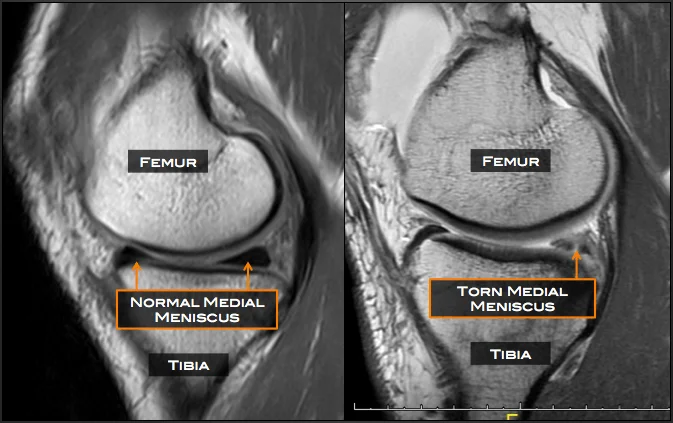
05. Meniscus Tears on MRI: Know the Patterns
Horizontal, Longitudinal & Radial Tears Explained
Instead of simply memorising the names of meniscal tears, understanding their orientation and MRI appearance makes them much easier to identify.
This session uses a simple banana analogy to explain the different tear patterns and their relationship with meniscal fibres.
🔑 Key Takeaways
- Horizontal meniscus tears
- Longitudinal vertical tears
- Radial tears
- MRI appearance of different tear patterns
- Circumferential fibres and hoop stress
- Meniscal extrusion
- Relationship between meniscal extrusion and osteoarthritis
- Important exam-oriented points
Quick Revision: What Did These Sessions Cover?
Before you move on, here’s your quick takeaway from all 5 sessions:
Session | Key Highlight |
Peripheral Artery Disease | PAD, imaging evaluation, Aortoiliac Occlusive Disease & limb ischemia |
Radiology Spotters | Cartagena Syndrome, GI Lymphoma, AVN, renal and pulmonary spotters |
FMF Preeclampsia Screening | FMF calculator, MoM, MAP, uterine artery Doppler & PAPP-A |
Fetal Hydrops | Umbilical vein/artery Doppler, absent/reversed EDF & fetal haemodynamics |
Meniscus Tears on MRI | Horizontal, longitudinal & radial tears, hoop stress & meniscal extrusion |
These are just the highlights. There’s much more to explore.
From Interventional Radiology and image-based spotters to fetal imaging and MRI, these sessions are designed to help you understand concepts rather than simply memorise them.
And remember — these 5 sessions are only a glimpse of what you can find inside Conceptual Radiology.
Want More High-Yield Radiology Learning?
Go beyond these highlights with the Conceptual Radiology platform, where you can access:
- Image-based learning
- Rapid revision videos
- High-yield spotters
- Solved DNB papers
- Live sessions
- Expert-led academic content
Don’t just scroll through the highlights. Watch the complete sessions and strengthen your concepts.
Download the eConceptual App and subscribe to Conceptual Radiology today.
See the image. Understand the concept. Revise smarter.